By: Maria Gemma del Remei Vercher Sifres
Obtaining a patent airway is crucial in daily practice to administer oxygen, inhalation drugs, or anesthetics for numerous interventions. For this purpose, there are both endotracheal and supraglottic intubation techniques, which avoid the insertion of devices into the tracheal lumen. However, in exotic animal practice, these procedures can be challenging as some patients have unique anatomical characteristics in their upper respiratory tract. Typically, their larynx and pharynx are smaller than in other species, and the long soft palate hinders the visualization of the epiglottis. Additionally, they have thick and fleshy lingual structures, long incisors, and large dental pieces.
Therefore, there are numerous intubation techniques described for each species studied, all of them with advantages and disadvantages that allow their routine use in exotic animal practice. Nevertheless, manufacturing and adapting existing tools and/or the innovative use of 3D printers can be considered to design and create instruments adapted to the unique anatomy of exotic animals, thus further facilitating the intubation procedure. Lastly, it’s essential to highlight that the success of this procedure also relies on minimal investment in both instruments and auxiliary aids, as well as the time dedicated to it, which depends on the veterinarian’s prior experience and training.
RABBIT – Oryctolagus cuniculus
These mammals have specific anatomical features, such as a long and narrow oropharynx inside a small oral cavity. They have limited temporomandibular joint mobility, a long tongue with an elevated fleshy base (lingual toruts), relatively large incisors for their size, a small glottis, and a larynx hidden behind the tongue.
For rabbit intubation, the most popular method is the blind technique, where the use of a laryngoscope is not necessary since the glottis of the patient is never seen. To verify correct tube placement, you can perform laryngeal palpation, observe the patient’s response in a shallow plane of anesthesia (coughing, gagging), monitor capnography results, and check for condensation inside the endotracheal tube and/or listen to the patient’s breathing through it.
However, intubation with direct visualization of the glottis can also be performed with the help of a simple laryngoscope, otoscope, or another illuminated speculum.
Finally, as alternative techniques, supraglottic airway devices (SGAD) have been described. In rabbits, the most studied is the V-gel as it is a species-specific device designed to adapt to anatomical structures and create a pressure seal in the pharynx. However, other devices like the laryngeal mask, laryngeal tube, or cobraPLA have also been used in human medicine.
GUINEA PIG – Cavia porcellus
Guinea pigs are obligate nasal breathers, and due to the palatal ostium, endotracheal intubation can be challenging. This small opening in the midline surface is the only connection between the oropharynx and the pharynx. Additionally, guinea pigs have a large and elongated tongue, which covers a significant portion of the mouth and oropharynx.
Since their intubation is so complicated, the best option is indirect visualization of the glottis using a guided endoscope, video laryngoscope, or video-optic stylet. Two techniques can be performed using these instruments: the “side by side” technique, where the endoscope remains in the oral cavity to visualize the larynx while the endotracheal tube is passed parallelly to reach the trachea, and the “over the endoscope” technique, in which the endoscope is inserted inside the endotracheal tube, and both instruments advance together toward the laryngeal opening.
COMMON RAT – Rattus norvegicus
The difficulty of the procedure primarily lies in the exposure and limited access to the laryngeal opening, which is small in rats (aditus laryngis) with a restricted range of oral opening. Like rabbits, rats have a long tongue with an elevated fleshy base (lingual tortus).
Due to the lack of materials adapted to the small size of this mammal, many methods and self-designed instruments are described to perform correct intubation. Thus, a pediatric laryngoscope blade can be used, or one can be fabricated by modifying an otoscope tube. Some studies have also employed the “side by side” technique as in guinea pigs. Other authors describe the fabrication of an intubation wedge made from a 3ml plastic syringe or an SGAD by modifying the design of the human laryngeal mask. There are even 3D-printed specific laryngoscope blades for rats.
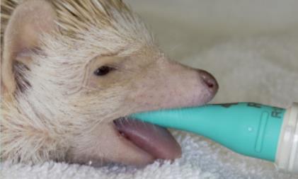
WHITE-BELLIED HEDGEHOG – Atelerix albiventris
Intubation in white-bellied hedgehogs is complicated due to their small size, a wide and voluminous tongue, prominent incisors, a small glottis, a narrow oropharynx, and large tonsils. The epiglottis is broad with a prominent apex, located dorsally to the soft palate, which must be gently displaced to visualize it. Additionally, this species is prone to developing multiple oral lesions that hinder oral cavity access, requiring resorting to tracheostomy.
To intubate these unique mammals, small endotracheal tubes or modified intravenous catheters can be used. Small-sized laryngoscopes can also be employed, and there are studies describing the successful use of the V-gel in hedgehogs.